Showing posts with label SCIENCE. Show all posts

Thursday, July 22, 2010

Saturday, May 22, 2010

The first artificial bacterial genome

Just about every science blogger in the world has written about J. Craig's working artificial bacterial genome. Most of them are much cleverer people than me, so I don't have much to say that wouldn't be repeated what has already been said, but I'd just like to point you to some posts that are good, or thought provoking, or hilarious.

Good:
Did scientists play god? 

Thought-provoking:
We can't control everything - evolution takes over immediately

Hilarious:
J. Craig versus Francis Collins

Just to add to that last one, I distinctly remember some lecturers in my undergrad talking about J. Craig Venter and emphatically adding that he's apparently a total asshole. Well, so what? I've met plenty of asshole scientists*; being a meanie isn't unique to JCV and it shouldn't take anything away from his achievements. And boy are they spectacular achievements.

*I would just like to make it clear that I am NOT referring to any of you lovely scientists who I have ever worked with. You guys rock.

Wednesday, January 20, 2010

Extraordinary Embryo of the Week


What a beauty! This is a mouse embryo, and you can tell that it has been stained for the expression of a gene. But this is not just any gene! This mouse has actually been genetically modified to contain some DNA from the extinct Tasmanian Tiger, Thylacinus cynocephalus.

Now, the point of this genetic modification wasn't to create a half-mouse-half-Tassie-tiger (that would just be absurd. Not to mention impossible...), but rather just to study the function of a particular gene. Because it would be damn near impossible to bring the Tasmanian tiger back from extinction, this approach is the best way to study its genetics.


The Tasmanian tiger

What the scientists did was very carefully extract DNA from four 100-year-old Tasmanian tiger specimens that had been preserved in alcohol, and amplify the DNA of interest (not an easy task if you're working with old DNA!). The DNA they amplified was from a region that controls the expression of a gene called Col2A1. You can think of this DNA as the 'switch' that turns Col2A1 on or off.

They attached the switch to an additional piece of DNA, a 'reporter' gene. Then, they inserted the whole DNA construct into a mouse genome. The reporter gene produces the blue pigment you can see. This method tells us where and when in the embryo the 'switch' is turning on. If the switch is turned on, the reporter gene is active and produces a blue pigment.

Basically, this was a really neat method for studying the function of a gene from an extinct animal! The blue pigment allows us to see where the gene is switched on, and then we can compare that to the mouse version of Col2A1. Turns out, Col2A1 seems to perform the same function whether it's from the Tasmanian tiger or the mouse (its function is in cartilage formation, which is why it is expressed in the forming bones).

This may not be a particularly thrilling conclusion, but the applications of the technique are pretty awesome. For example, maybe one day we could examine what dinosaurs looked like, if we could extract the relevant genes from dinosaurs and insert them into another animal!

And actually, geneticists use this technique for non-extinct animals as well. It's a really good way to figure out if a similar gene performs the same function in different animals. These kinds of studies tell us about the evolutionary history of individual genes, which is bloody interesting, if you ask me.

Reference: Pask, A.J., Behringer, R.R., Renfree, M.B., 2008. Resurrection of DNA Function In Vivo From an Extinct Genome. PLoS ONE, 3(5), e2240.

Tuesday, January 5, 2010

Extraordinary Embryo of the Week

Oops, a day late again. I was shopping yesterday. I'm sure all 2 of my readers understand that me getting a bargain price on some beautiful black patent leather pumps is worth a slightly late blog post :D

Anyway:


This little guy is a Xenopus laevis embryo that I stained for the expression of a gene called FGF-8 (fibroblast growth factor 8).

FGF-8 is important for the development of many different tissues throughout the body - you can see at this stage that FGF-8 is turned on in the tail bud (to the right of the picture), the somites (the stripy bits along its middle), the midbrain-hindbrain boundary (the stripe at the top of its head) and a couple of the branchial/pharyngeal arches (other stripy bits on its head).

So what?

This gene is being turned on in a bunch of different places. And in each different tissue, it is doing a slightly different job. How can the same gene have different functions in different places?

It all depends on context. The environment that the cells are in, and the complement of genes that are turned on in each cell, all affect how FGF-8 functions. It's kind of a space-saver in the genome; instead of having a different set of genes for every conceivable developmental job, we find that some genes are re-used all over the body to control the development of different organs and tissues, and FGF-8 is just one of these multifunctional genes.

FGF-8 does even more work during development at different developmental stages. For example, later on in development, FGF-8 will be used to control the growth of the developing limbs.

So, our Xenopus embryo above illustrates a couple of really fundamental ideas in developmental biology:

1. The same gene can perform different functions in various tissues (AND at different time-points).

2. The environment and genetic context affect how a gene will function.

I'd really like to pause here, and I'll pick up later this week to explore what these points mean for the evolution of developmental systems.

Monday, December 28, 2009

Extraordinary Embryo of the Week

OK. So I have two weeks of embryos to catch up on... blame the Christmas madness for my neglect of the blog!

First up, a sea urchin embryo, from George Watchmaker at Livermore, CA, USA.


Sea urchins were one of the first model systems in developmental biology, and the first species in which sperm cells were shown to fertilise the ovum! Nowadays they're often used by groups doing evolution and development (evo-devo) studies.

Now for the second picture: this time, it's a human!


This embryo is just starting to grow limbs, you can see one of the limb buds as a big blob coming off the side of the embryo. Even now, when it looks like a shapeless blob, the limb already has all three axes determined: dorsal-ventral (back to palm of hand), anterior-posterior (thumb to pinky - even though the digits haven't yet formed!) and proximal-distal (shoulder to fingertip). I'll write a more detailed post on limb development (and what can go wrong) sometime in the future, because it's a really great example of organ development in an embryo.


I hope everyone had a great Christmas! I can't believe it's almost 2010. I have to start thinking about my New Year's resolutions... eek

Saturday, December 12, 2009

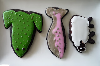
Science cookies

Inspired by these beautiful cookies, my friends and I made a bunch of geeky science-themed cookies today!

Here is one of our (far inferior) electrophoresis gel cookies...


And then we got a little carried away with other geeky things too...


Including the animals we work on: Xenopus laevis, Oncorhynchus tshawytscha, and Ovis aries


Sequencing electropherograms: We designed sequences that spelt out our names in one-letter amino acid codes!


And little models of cells that include features such as a nucleus, rough ER and smooth ER, mitochondria, and Golgi.


I don't think I'll need to eat for a month after all these...

Monday, December 7, 2009

We are made of star stuff

I'm currently reading the very cool book Death From The Skies!, by Phil Plait, which outlines a bunch of end-of-the-world scenarios and then talks about the science behind them. Phil's an astronomer, and a fantastic writer - I've been a fan of his ever since I was about 15 years old, when I discovered his hilarious Bad Astronomy website (his review of the movie Armageddon still makes me chuckle).

Death From The Skies! is one of those books where you just end up thinking "wow, science is so darn cool!". Being a bit of a geek for pretty much my entire life, I thought I knew a reasonable amount about astronomy and astrophysics, for someone who's a non-specialist. But every chapter of Death From The Skies is full of things that I haven't encountered before - and many of these tidbits are extremely cool.

One particular chapter on supernovae is full of "wow-facts". In this chapter, Phil talks about the processes of fusion in the core of massive stars. Our own sun fuses hydrogen nuclei to produce helium, but more massive stars can produce other elements by fusion, once their hydrogen supply has run out. Massive stars produce, in this order, carbon, neon, oxygen and silicon; and the most massive stars in our universe have such tremendous pressure and heat at their core that they can produce iron. These stars, however, do not have long to live once they are producing iron, and will soon explode in a furious cascade of events that we call a supernova.

The early universe contained only three elements: hydrogen, helium, and a little bit of lithium. Nothing else. Then supermassive stars started to form, and began creating heavier elements from the lighter ones, right up to iron, then exploded. The explosions of these stars triggered the formation of new stars... and the cycle continued.

I'll leave you with (what I think is) the most striking paragraph of this chapter:

When you cut your finger and a thin rivulet of blood seeps up into the slice, the red color you see is due to hemoglobin, and the key factor in that molecule is iron. That iron was forged in the heart of a supernova.

As Carl Sagan told us: We're made of star stuff.


Tuesday, October 27, 2009

Axolotl development

In our department we have a breeding pair of axolotls in the teaching labs. They've had some babies recently, and the baby axolotls have just hatched from their eggs. They're awfully cute, they look quite similar to Xenopus tadpoles except they've got those fantastic external gills.

I looked up a table of axolotl development and found a site with an excellent series of photographs, depicting the development right through to larval stage. The Late Neurula stage is SO COOL. Go look at it. Now.