Showing posts with label Extraordinary Embryo of the Week. Show all posts

Wednesday, January 20, 2010

Extraordinary Embryo of the Week


What a beauty! This is a mouse embryo, and you can tell that it has been stained for the expression of a gene. But this is not just any gene! This mouse has actually been genetically modified to contain some DNA from the extinct Tasmanian Tiger, Thylacinus cynocephalus.

Now, the point of this genetic modification wasn't to create a half-mouse-half-Tassie-tiger (that would just be absurd. Not to mention impossible...), but rather just to study the function of a particular gene. Because it would be damn near impossible to bring the Tasmanian tiger back from extinction, this approach is the best way to study its genetics.


The Tasmanian tiger

What the scientists did was very carefully extract DNA from four 100-year-old Tasmanian tiger specimens that had been preserved in alcohol, and amplify the DNA of interest (not an easy task if you're working with old DNA!). The DNA they amplified was from a region that controls the expression of a gene called Col2A1. You can think of this DNA as the 'switch' that turns Col2A1 on or off.

They attached the switch to an additional piece of DNA, a 'reporter' gene. Then, they inserted the whole DNA construct into a mouse genome. The reporter gene produces the blue pigment you can see. This method tells us where and when in the embryo the 'switch' is turning on. If the switch is turned on, the reporter gene is active and produces a blue pigment.

Basically, this was a really neat method for studying the function of a gene from an extinct animal! The blue pigment allows us to see where the gene is switched on, and then we can compare that to the mouse version of Col2A1. Turns out, Col2A1 seems to perform the same function whether it's from the Tasmanian tiger or the mouse (its function is in cartilage formation, which is why it is expressed in the forming bones).

This may not be a particularly thrilling conclusion, but the applications of the technique are pretty awesome. For example, maybe one day we could examine what dinosaurs looked like, if we could extract the relevant genes from dinosaurs and insert them into another animal!

And actually, geneticists use this technique for non-extinct animals as well. It's a really good way to figure out if a similar gene performs the same function in different animals. These kinds of studies tell us about the evolutionary history of individual genes, which is bloody interesting, if you ask me.

Reference: Pask, A.J., Behringer, R.R., Renfree, M.B., 2008. Resurrection of DNA Function In Vivo From an Extinct Genome. PLoS ONE, 3(5), e2240.

Tuesday, January 5, 2010

Extraordinary Embryo of the Week

Oops, a day late again. I was shopping yesterday. I'm sure all 2 of my readers understand that me getting a bargain price on some beautiful black patent leather pumps is worth a slightly late blog post :D

Anyway:


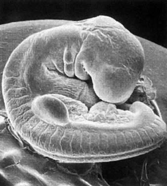
This little guy is a Xenopus laevis embryo that I stained for the expression of a gene called FGF-8 (fibroblast growth factor 8).

FGF-8 is important for the development of many different tissues throughout the body - you can see at this stage that FGF-8 is turned on in the tail bud (to the right of the picture), the somites (the stripy bits along its middle), the midbrain-hindbrain boundary (the stripe at the top of its head) and a couple of the branchial/pharyngeal arches (other stripy bits on its head).

So what?

This gene is being turned on in a bunch of different places. And in each different tissue, it is doing a slightly different job. How can the same gene have different functions in different places?

It all depends on context. The environment that the cells are in, and the complement of genes that are turned on in each cell, all affect how FGF-8 functions. It's kind of a space-saver in the genome; instead of having a different set of genes for every conceivable developmental job, we find that some genes are re-used all over the body to control the development of different organs and tissues, and FGF-8 is just one of these multifunctional genes.

FGF-8 does even more work during development at different developmental stages. For example, later on in development, FGF-8 will be used to control the growth of the developing limbs.

So, our Xenopus embryo above illustrates a couple of really fundamental ideas in developmental biology:

1. The same gene can perform different functions in various tissues (AND at different time-points).

2. The environment and genetic context affect how a gene will function.

I'd really like to pause here, and I'll pick up later this week to explore what these points mean for the evolution of developmental systems.

Monday, December 28, 2009

Extraordinary Embryo of the Week

OK. So I have two weeks of embryos to catch up on... blame the Christmas madness for my neglect of the blog!

First up, a sea urchin embryo, from George Watchmaker at Livermore, CA, USA.


Sea urchins were one of the first model systems in developmental biology, and the first species in which sperm cells were shown to fertilise the ovum! Nowadays they're often used by groups doing evolution and development (evo-devo) studies.

Now for the second picture: this time, it's a human!


This embryo is just starting to grow limbs, you can see one of the limb buds as a big blob coming off the side of the embryo. Even now, when it looks like a shapeless blob, the limb already has all three axes determined: dorsal-ventral (back to palm of hand), anterior-posterior (thumb to pinky - even though the digits haven't yet formed!) and proximal-distal (shoulder to fingertip). I'll write a more detailed post on limb development (and what can go wrong) sometime in the future, because it's a really great example of organ development in an embryo.


I hope everyone had a great Christmas! I can't believe it's almost 2010. I have to start thinking about my New Year's resolutions... eek

Monday, December 14, 2009

Extraordinary Embryo of the Week

Mondays are tough. It seems to be a universal truth.

My solution? Brighten our Mondays with a beautiful embryo image!

Of course, for my first Extraordinary Embryo, I had to choose Xenopus laevis, my current weapon of choice (slash model organism).

I've had this image on my screensaver for a while now, and while it's very simple, it's also quite beautiful. It's from Michael Klymkowsky at the University of Colorado Boulder.


As you can probably tell, they're both quite early stage embryos - they're still just balls of cells. The embryo on the left is at a later stage than the one on the right - you can see that it has a lot more cells.

There's something about these ball-shaped embryos that really makes me smile. Why? I guess because they make me think about what's still to come.

At some point they stop being balls and start to become animals. The cells in these balls move and grow and change, and they do so in an organised way. Cells on one side "know" that they're on that side, and act accordingly. Every cell is an individual, there is no "boss-cell" giving orders and telling the rest where to go. Yet somehow, these individuals work together, dividing and differentiating and migrating... To produce an animal.

And I think that's pretty amazing.